
Citrus Micrografting for Regrowth After Shoot Tip Cryopreservation
Gayle M. Volk, USDA-ARS National Laboratory for Genetic Resources Preservation, 1111 S. Mason St., Fort Collins, Colorado 80521. Gayle.Volk@usda.gov
Remi Bonnart, USDA-ARS National Laboratory for Genetic Resources Preservation, 1111 S. Mason St., Fort Collins, Colorado 80521.
Ashley Shepherd, USDA-ARS National Laboratory for Genetic Resources Preservation, 1111 S. Mason St., Fort Collins, Colorado 80521.
Outline
- Introduction
- Prepare the rootstock seedlings
- Warm the cryopreserved shoot tips
- Micrograft the shoot tips onto the rootstocks
- Shoot tip regrowth and trimming
- References
- Associated information
- Acknowledgments
The citrus micrografting procedure is available to download here.
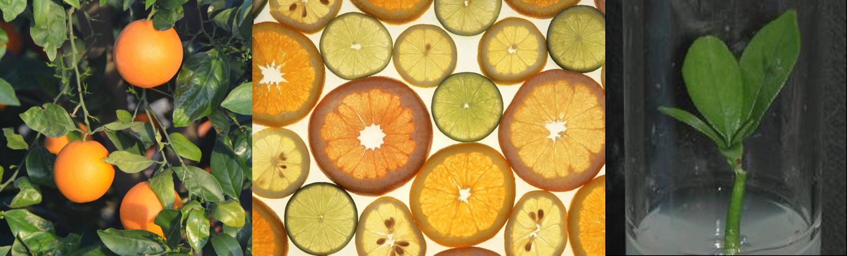
1. Introduction
This chapter demonstrates the process of micrografting as a method to recover citrus shoot tips after cryopreservation. Micrografting is a process whereby shoot tips are attached to seedling rootstocks and then grow into healthy plants. Micrografting is used to recover citrus shoot tips because they failed to grow when they were placed directly on medium, likely because the medium was not optimized for their growth requirements.
The micrografting method is demonstrated as a series of steps including:
- Preparing the seeds to produce rootstock seedlings
- Warming the cryopreserved citrus shoot tips
- Trimming the cryopreserved citrus shoot tips and micrografting them onto the seedling rootstocks
- Regrowth of the micrografted plants
2. Prepare the rootstock seedlings
‘Carrizo’ citrange rootstock seeds (PI 150916 x Citroncirus sp.) were selected as the rootstock cultivar because they are available from nursery sources, they produce relatively thick hypocotyls for grafting, and they are compatible with nearly every species of citrus.
Video 1 demonstrates the process of peeling the seed coats off the ‘Carrizo’ seeds and placing the resulting embryos into water containing Tween 20 (1 drop per 100 mL). When all the seed coats are removed, the seeds are rinsed three times with tap water and then surface sterilized by first treating the embryos with 70% isopropanol for 2 minutes and then rinsed three times with tap water. 20% bleach (1.65% sodium hypochlorite final concentration) + Tween 20 (1 drop per 100 mL) is then added to the flask and the embryos are transferred to a laminar flow hood. The seeds are shaken intermittently for 20 minutes while they are in the bleach solution. They are then rinsed three times with sterile water and embryos are transferred to test tubes containing the growth medium Citrus Seed Germination Medium. The embryos in the test tubes are then placed in a dark cabinet in a growth room and grown for up to six weeks before use.
Video 1. USDA-ARS Technician Remi Bonnart demonstrates the seed coat removal, surface sterilization, and introduction onto medium processes.
3. Warm the cryopreserved shoot tips
When the rootstocks are at least 3 cm tall, they are ready to be micrografted. Shoot tips that were previously cryopreserved (see the “Citrus shoot tip cryopreservation” chapter) are recovered from liquid nitrogen and warmed.
First, the cryovial is removed from the cryotank in which it has been stored in liquid nitrogen (LN) vapor. Cryovials are stored on cryocanes in cryoboxes in the tank. The vial of interest in quickly removed from the cryocane and placed into LN and transferred to a dewar with LN in the laminar flow hood. This process is shown in Video 2.
Video 2. USDA-ARS Technician Remi Bonnart retrieves a cryovial from long-term storage in the vapor-phase of LN.
Within the laminar flow hood, the cryovial is opened and the aluminum foil strip containing the shoot tips is quickly transferred to a room-temperature (22 oC) Unloading Solution (1.2 M sucrose + 1/2 strength MS). The shoot tips are incubated in the unloading solution for 20 minutes at 22 oC. The solution is then removed and shoot tips are plated onto Citrus Shoot Tip Recovery Medium (Video 3). They remain on the medium for 18 hours at 25 oC in the dark before they are micrografted onto rootstocks.
Video 3. USDA-ARS Technician Remi Bonnart demonstrates the shoot tip warming process.
4. Micrograft the shoot tips onto the rootstocks
The dissecting microscope is placed in a laminar flow hood and the stage is surface sterilized with 70% ethanol. Rootstocks are then retrieved from the test tubes and the root is trimmed to a length of 3 to 4 cm (Fig. 1). The hypocotyl is cut at a height of about 1 cm above the root (Fig. 1). The rootstock is prepared for micrografting by carefully cutting a small ledge on the hypocotyl, with a 2 mm deep incision made to bisect the cut surface, followed by a perpendicular cut from the edge of the seedling. Approximately 0.2 mm of the basal portion of the shoot tip is removed, and the shoot tip is then placed onto the rootstock ledge (Fig. 2). If needed, shoot tips are moistened with Shoot Tip Dip/Rinse Solution (3% sucrose + MS solution). The seedling rootstock with the shoot tip is then placed onto Citrus Micrograft Recovery Medium in 25 x 150 mm test tubes with 25 mL of medium per tube. The plant cultures are grown at 25 oC with ~100 µmol m-2 s-1 light provided by fluorescent bulbs with a 16 h photoperiod. Video 4 demonstrates these steps of the procedure.
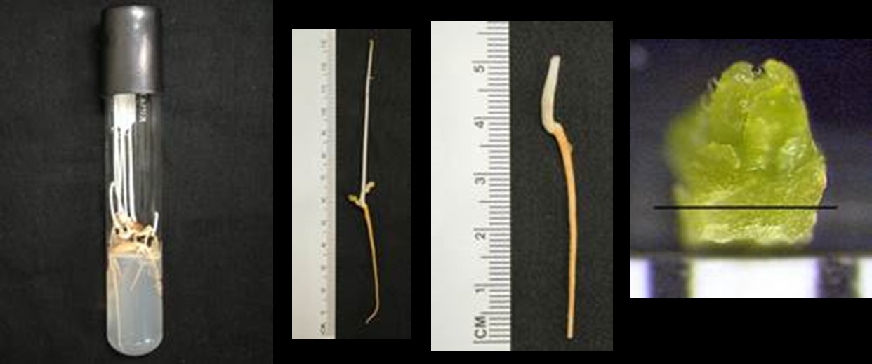
Figure 1. Seedling rootstocks in a test tube ready to be used for micrografting (left). Seedling rootstock before and after trimming (middle). Shoot tip close-up with a line showing where the basal portion of the shoot tip will be trimmed prior to micrografting. Photo credit: Ashley Shepherd.
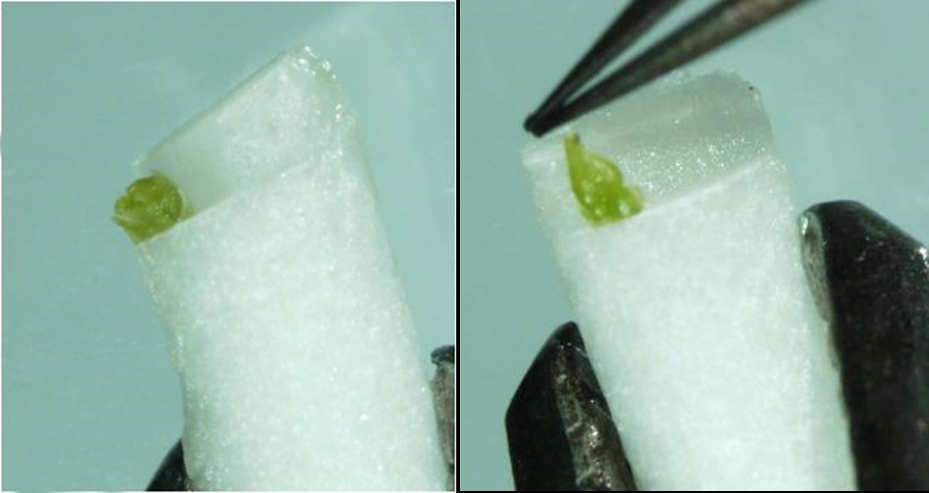
Figure 2. Trimmed shoot tip placed on the rootstock ledge during the micrografting process. Photo credit: Remi Bonnart.
Video 4. USDA-ARS Technician Ashley Shepherd demonstrates the rootstock trimming and shoot tip micrografting process (narrated by Remi Bonnart).
5. Shoot tip regrowth and trimming
Micrografted plants are grown for several weeks and shoot tip regrowth will be visible. In some cases, lateral shoots grow from the rootstock. Those non-shoot tip derived shoots must be removed so that they do not compete with the elongating shoot tip and inhibit its growth. The following video (Video 5) and images (Fig. 3 and 4) shows citrus plants recovered 6 to 8 weeks after shoot tips have been micrografted. If lateral shoots are apparent, they are removed and the plants are returned to the test tubes, which is also demonstrated in the video. Regrowth data are usually collected after 6 to 8 weeks. Micrografted citrus plants may then be transferred and grown on soil (Fig. 5 and 6).
Video 5. USDA-ARS Technician Ashley Shepherd shows citrus plants recovered after micrografting the cryopreserved shoot tips and the process of trimming shoots that may grow directly from the rootstock and did not originate from the micrografted shoot tip (narrated by Remi Bonnart).
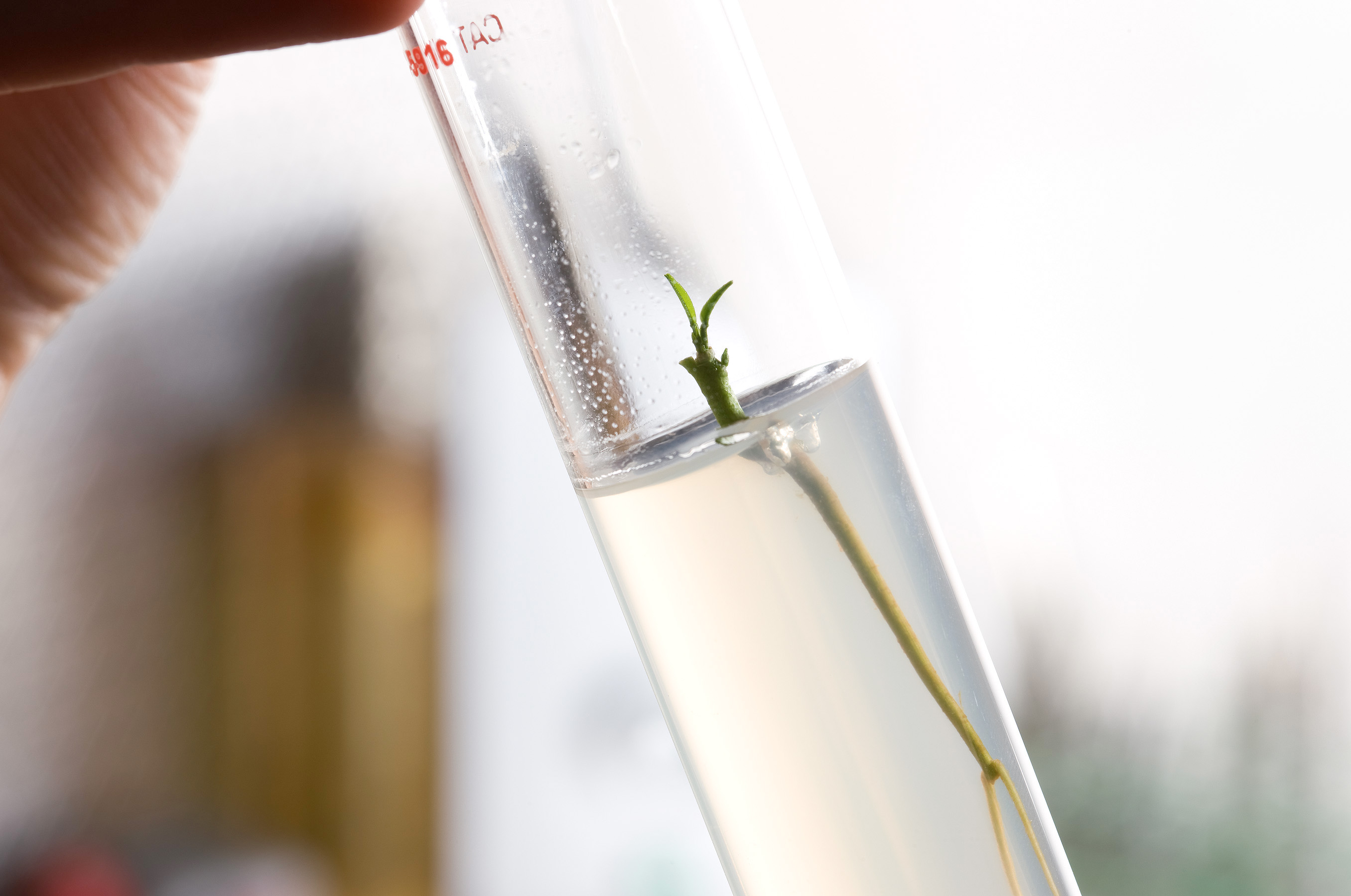
Figure 3. Micrografted citrus shoot tip that has regrown into a plant. Photo credit: Peggy Greb.

Figure 4. Shoot tips that have recovered into plants that could be transferred to the greenhouse. Photo credit: Gayle Volk.
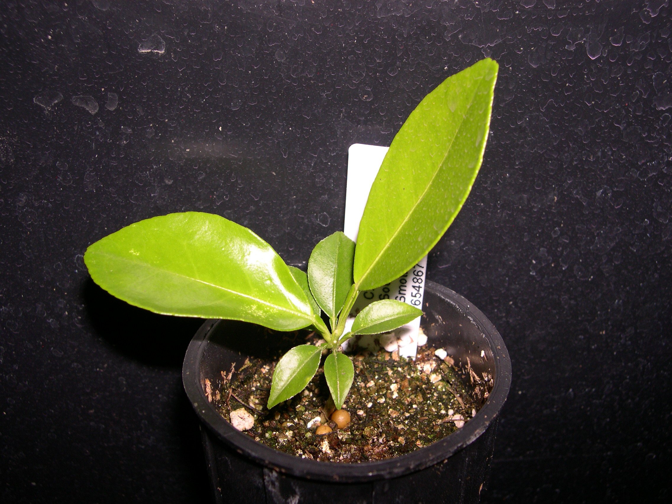
Figure 5. Micrografted citrus plant that has been been transferred to soil and grown in the greenhouse. Photo credit: Remi Bonnart.
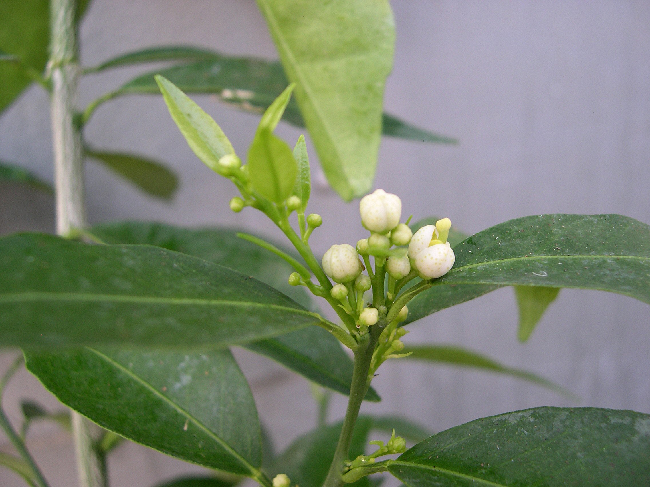
Figure 6. Citrus plant in the greenhouse derived from cryopreserved shoot tips. The resulting citrus plants flower within 12 to 15 months. Photo credit: Remi Bonnart.
6. References
Volk, GM, Bonnart R, Krueger R, Lee R. 2012. Cryopreservation of Citrus shoot tips using micrografting for recovery. CryoLetters 33:418-426.
Volk GM, Bonnart R, Shepherd A, Krueger R, Lee R. 2015. Cryopreservation of Citrus for Long-term Conservation. Acta Hort. 1065:187-192.
Volk GM, Bonnart R, Shepherd A, Yin Z, Lee R, Polek ML, Krueger R. 2016. Citrus cryopreservation: Viability of diverse taxa and histological observations. Plant Cell Tissue and Organ Culture 128:327-334. DOI 10.1007/s11240-016-1112-4
Volk GM, Jenderek MM, Walters C, Bonnart R, Shepherd A, Skogerboe D, Hall BD, Moreland B, Krueger R, Polek ML. 2019. Implementation of Citrus shoot tip cryopreservation in the USDA-ARS National Plant Germplasm System. Acta Hort. 1234: 329-334.
7. Associated information
The citrus micrografting procedure is available to download here.
A large-scale citrus cryopreservation effort took place at NLGRP in 2016-2017. Reports were provided on the monthly progress of the project. A compilation of the monthly reports is available here.
8. Acknowledgments
Citation: Volk GM, Bonnart R, Shepherd A. 2020. Citrus Micrografting for Regrowth After Shoot Tip Cryopreservation. In: Volk GM (Eds.) Training in Plant Genetic Resources: Cryopreservation of Clonal Propagules. Fort Collins, Colorado: Colorado State University. Date accessed. Available from https://colostate.pressbooks.pub/clonalcryopreservation/chapter/citrus-micrografting/
This training module was made possible by:
Editors: Emma Balunek, Gayle Volk
Content providers: Remi Bonnart, Ashley Shepherd, Peggy Greb
Videographers: Mike May, Remi Bonnart, Gayle Volk
USDA is an equal opportunity provider, employer, and lender. Mention of trade names or commercial products in this article is solely for the purpose of providing specific information and does not imply recommendation or endorsement by the U.S. Department of Agriculture.
